MyPath Melanoma is a 23-gene expression profile test that provides objective information to aid in the diagnosis of challenging melanocytic lesions. MyPath Melanoma has a higher sensitivity than PRAME IHC, resulting in far fewer false negatives (97% vs. 59%).
Performance of PRAME staining in ambiguous melanocytic lesions
PRAME IHC can be useful, but there are many melanomas that are PRAME negative and some non-melanomas that are PRAME positive. Additionally, many times PRAME can show partial positivity or even weak positivity which is inconclusive.
- Inconclusive staining (1+, 2+, 3+) occurs in both nevi and melanomas and thus is unlikely to add value in achieving a definitive diagnosis.1
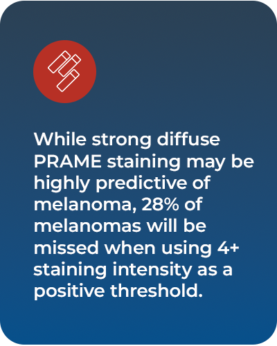
- In a meta-analysis, at a threshold of 4+ for diagnosing melanoma, PRAME has a sensitivity of 71% and specificity of 98%. While this results in a high positive predictive value, 28% of melanomas will be missed.2
- Performance is variable by subtype with lower sensitivity in desmoplastic (35%)1 and spitzoid (28%)2 lesions.

Association of PRAME IHC and GEP in challenging cutaneous melanocytic lesions
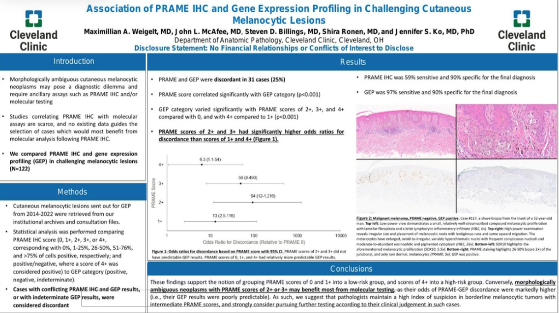
A recent study from Cleveland Clinic found GEP testing to have a sensitivity of 97% compared to PRAME IHC with a sensitivity of 59% in the same cohort of lesions. The study also highlighted the usefulness of GEP (MyPath Melanoma) in inconclusive PRAME IHC results (especially 2+ and 3+).

SUMMARY
- PRAME staining patterns were compared to GEP results in 122 challenging melanocytic lesions.
- PRAME IHC at a 4+ positive cut-off was 59% sensitive and 90% specific.
- MyPath Melanoma GEP was 97% sensitive and 90% specific.
- PRAME scores of 2+ and 3+ were more likely to be discordant with GEP results than scores of 4+.
CONCLUSIONS
- GEP has a notably higher sensitivity than PRAME IHC staining resulting in far fewer false negatives.
- Pathologists must maintain a high level of suspicion in borderline melanocytic tumors with intermediate PRAME scores and strongly consider pursuing further testing, especially for those with scores of 2+ or 3+ which may benefit the most from molecular testing.

